Latest Technology at Mark Hinds Optometrists
At Mark Hinds Optometrists, we invest heavily in advanced diagnostic and imaging technology to provide patients with highly detailed, accurate and personalised eye care.
Our equipment allows us to assess not only your vision, but also the health, structure and function of the eye — from the tear film and cornea through to the retina, optic nerve and visual field. This helps us detect disease earlier, monitor change more precisely, and tailor treatment to each patient’s needs.
Comprehensive Vision Testing
Across multiple consultation rooms, our practice is equipped with advanced visual acuity and refraction technology, including:
- Topcon TRK-2P auto-refractor
- Auto-keratometry
- Auto-pachometry for central corneal thickness measurement
- CV-5000 computerised refractor head and vision testing system for precise spectacle and vision assessment
We also use multiple methods of intraocular pressure measurement, including:
- Non-contact tonometry
- Goldmann applanation tonometry
- Icare tonometry
- Corvis ST biomechanically corrected IOP measurement
This allows us to select the most appropriate and accurate method depending on the patient, the condition being monitored, and the clinical context.
Advanced Corneal, Keratoconus and Anterior Eye Imaging
See our blog on how we successfully fit complex corneas.
Mark Hinds Optometrists uses state-of-the-art anterior segment imaging for corneal disease, contact lens fitting, keratoconus monitoring and surgical co-management.
Our technology includes:
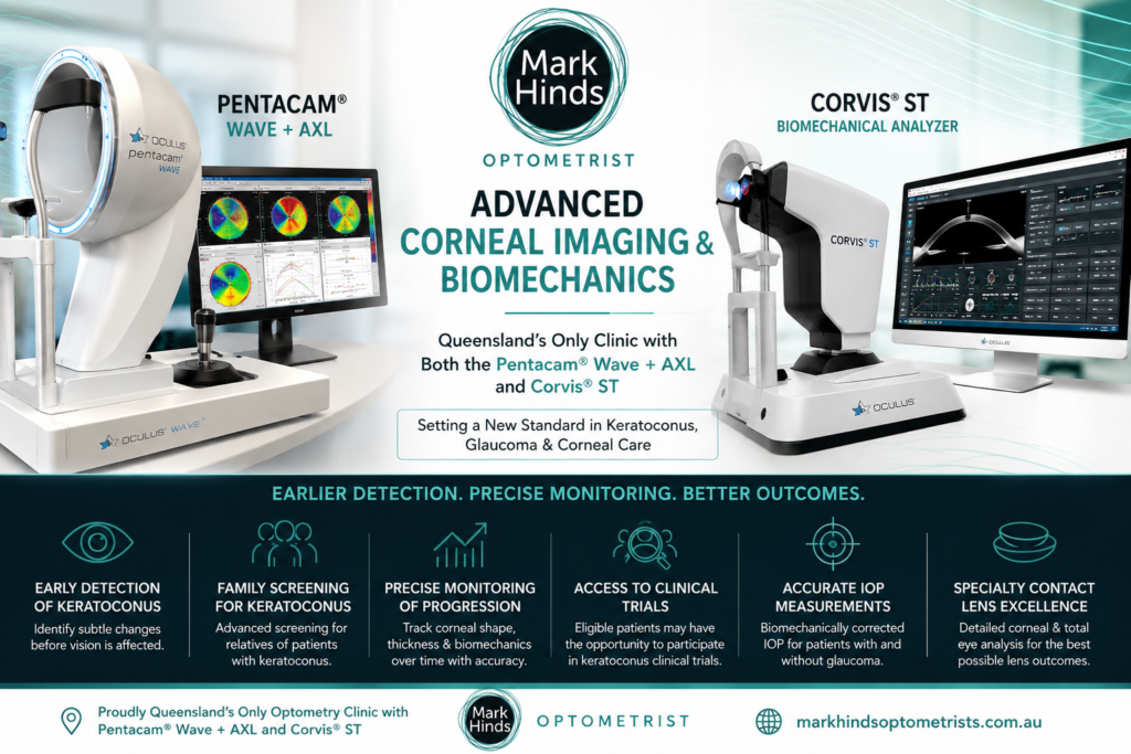
OCULUS Pentacam® AXL Wave
The Pentacam AXL Wave provides advanced anterior eye segment tomography, axial length measurement and wavefront analysis. It is particularly valuable for:
- Keratoconus detection and monitoring
- Corneal shape analysis
- Myopia management and axial length monitoring
- Pre- and post-surgical assessment
- Complex contact lens planning
- Anterior chamber and corneal thickness evaluation
OCULUS Corvis® ST
The Corvis ST provides a unique assessment of corneal biomechanics and biomechanically corrected intraocular pressure. It helps us better understand the strength, behaviour and risk profile of the cornea, particularly in patients with keratoconus, glaucoma risk, ocular hypertension or complex corneal disease.
Medmont Meridia and OCULUS Keratograph
Our advanced corneal topography systems allow us to map the front surface of the eye in detail. The Medmont Meridia also provides:
- Infrared meibomian gland imaging
- Fluorescein imaging
- Tear film analysis
- Digital contact lens simulation
- Imaging that can be shared directly with laboratories for custom lens design
sMap3D Scleral Profilometry
For advanced scleral and specialty contact lens fitting, we use sMap3D profilometry to capture the shape of the sclera and create highly customised scleral lens designs. This is especially useful for patients with:
- Keratoconus
- Post-corneal graft eyes
- Irregular corneas
- Ocular surface disease
- Complex contact lens fitting needs
Specular Microscopy for Corneal Health
Our Topcon Specular Microscope allows us to measure endothelial cell density and assess the health of the innermost layer of the cornea.
This is particularly important for patients with conditions such as Fuchs’ endothelial dystrophy, corneal disease, previous eye surgery, and for safety monitoring in clinical trial research.
Digital Retinal Imaging
For digital retinal imaging, we use Topcon’s advanced non-mydriatic retinal camera technology, including the Topcon NW500 Non-Mydriatic Retinal Camera.
This provides high-quality retinal images, often without requiring dilation, and is especially useful for patients with smaller pupils or those requiring ongoing ocular health monitoring.
Digital retinal imaging helps us assess and monitor conditions such as:
- Diabetic eye disease
- Macular degeneration
- Glaucoma
- Retinal vascular disease
- Optic nerve abnormalities
- General retinal health changes over time
Optical Coherence Tomography and OCT Angiography
Optical Coherence Tomography, or OCT, provides highly detailed cross-sectional imaging of the eye. It allows us to assess fine structural changes in the retina, macula, optic nerve and anterior segment.
OCT is an essential tool for the detection, monitoring and management of:
- Age-related macular degeneration
- Glaucoma
- Diabetic eye disease
- Optic nerve disease
- Macular disease
- Vitreoretinal interface disorders
- Keratoconus and anterior segment abnormalities
- Scleral and rigid contact lens fitting
Our OCT technology also includes OCT Angiography, which images the retinal blood vessel layers without dye injection. This is particularly valuable for patients at risk of choroidal neovascular membranes, diabetic eye disease and macular degeneration.
Glaucoma and Macular Degeneration Monitoring
Our glaucoma patients are carefully monitored using a combination of intraocular pressure measurement, optic nerve imaging, OCT retinal nerve fibre layer analysis, visual field testing and clinical examination.
Where required, patients are co-managed with specialist ophthalmologists to ensure optimal treatment and long-term care.
For patients with age-related macular degeneration, OCT and retinal imaging allow us to monitor for changes over time. This is especially important in patients with dry macular degeneration, where regular scans can help detect early conversion to wet macular degeneration.
Visual Field Testing
We use multiple Medmont Visual Field analysers with video monitoring to accurately assess and monitor visual field loss.
Visual field testing is important in the diagnosis and management of:
- Glaucoma
- Optic nerve disease
- Neurological visual pathway defects
- Retinal disease
- Macular disease
- Unexplained vision loss
Myopia Control Clinic
Our paediatric myopia control clinic uses advanced technology to monitor eye growth and myopia progression.
This includes axial length measurement with the Pentacam AXL Wave and OCT biometry capability. Axial length measurement is an important part of modern myopia management because it helps us monitor the physical growth of the eye, not just changes in spectacle prescription.
This technology supports evidence-based treatment options such as:
- Orthokeratology
- Myopia control contact lenses
- Myopia control spectacle lenses
- Atropine therapy where clinically indicated
- Ongoing progression monitoring
Specialty Contact Lens Technology
Mark Hinds Optometrists has an extensive range of trial contact lenses, including soft, rigid gas permeable, hybrid, mini-scleral and scleral lenses.
We regularly adopt new contact lens technologies, including pre-market and emerging lens designs where available through research and professional collaboration.
Our specialty contact lens technology assists patients with:
- Keratoconus
- Post-graft corneas
- Irregular astigmatism
- High prescriptions
- Dry eye disease
- Ocular surface disease
- Complex visual needs
- Failed or limited success with standard contact lenses
Clinical Trial and Research Infrastructure
Helping others through research and clinical trials.
Many of our patients have also been involved in clinical research through Dr Mark Hinds’ clinical trial practice, Ophthalmic Trials Australia.
This research arm allows suitable patients to access emerging technologies, treatments and contact lens innovations before they become widely available.
Our practice also includes research-grade infrastructure such as:
- Temperature-controlled and remotely monitored 2–8°C fridges
- -20°C freezers
- Backup power and generator support
- Designated pharmacy facilities
- Vital sign monitoring equipment including blood pressure, ECG, SpO₂ and tympanic temperature measurement
This infrastructure supports our role in ophthalmic clinical trials and reflects our commitment to advancing eye care.
Access to Emerging Treatments
For patients whose current treatments are not suitable or who have complex eye disease, clinical trial participation may sometimes be an option through Ophthalmic Trials Australia.
Clinical trials allow eligible participants to access innovative therapies that are not yet available to the general public, while contributing to the future of eye care.
At Mark Hinds Optometrists, our goal is simple: to combine clinical experience, advanced technology and research-driven care to provide the most comprehensive eye care possible.
Check us out at ot-au.com to see some of our research in publication: