What happens over the day
Immediately after insertion, the reservoir is mostly preservative-free filling solution mixed with some basal tears. During wear, several things occur:
- Reservoir thickness reduces
The lens settles into the conjunctiva/sclera, so the post-lens fluid reservoir becomes thinner, especially in the first 1–2 hours. A 2025 OCT study found the fluid reservoir thickness decreased significantly over 4 hours of scleral lens wear. - Tear exchange is low and variable
Some lenses allow a little tear inflow/mixing; others behave almost like a sealed system. The literature generally supports that tear exchange is minimal compared with normal blinking, so substances can accumulate under the lens. - Debris and inflammatory mediators can accumulate
This is the basis of midday fogging. Particulate matter may include epithelial cells, inflammatory cells, lipid, mucin-like material, protein, and tear debris. Midday fogging has been reported in roughly 26–46% of scleral lens wearers in some studies.
In one study, MMP-9 and MMP-10 were higher in the scleral lens fluid reservoir than in basal tears after wear, supporting the idea that the reservoir can become a relatively stagnant inflammatory microenvironment.
Does pH change?
Probably yes, but direct human data measuring reservoir pH over time is limited.
Mechanistically, the post-lens reservoir can drift more acidic because:
- the cornea continues to metabolise glucose;
- relative hypoxia promotes more anaerobic metabolism;
- lactate and CO₂ accumulate;
- CO₂ retention shifts the bicarbonate buffer system toward lower pH;
- tear exchange may be insufficient to fully refresh the compartment.
So the concern is not just “saline under a lens”; it is a semi-closed metabolic space. Contact lens wear can contribute to corneal acidosis through hypoxia and CO₂ accumulation, and this is conceptually relevant to scleral lenses because the lens/reservoir complex slows gas and solute exchange.
Clinically, this is why buffered, preservative-free scleral filling solutions are often better tolerated than plain unbuffered saline. Commercial scleral salines may range from about pH 5.3 to 7.4, whereas physiologic tears are closer to neutral/slightly alkaline.
My practical interpretation:
pH likely trends downward during wear in a poorly exchanging reservoir, especially with thick vault, low Dk/t, tight landing, inflammatory surface disease, or unbuffered saline. Buffered electrolyte solutions reduce the initial pH mismatch but do not completely prevent metabolic change over a long wearing day.
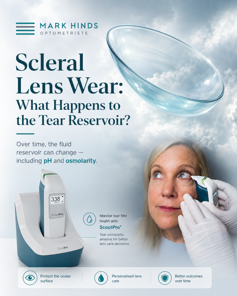
Does osmolarity change?
Yes, but it depends what you are measuring: the exposed tear film versus the post-lens reservoir.
Exposed / pre-lens tear osmolarity
Scleral lenses often reduce tear evaporation and protect the ocular surface. In keratoconus patients, Carracedo et al. found tear osmolarity decreased after 6–9 hours of scleral lens wear, while MMP-9 increased.
That makes sense clinically: a scleral lens can improve the dry-eye environment by shielding the cornea from evaporation and desiccation.
Post-lens reservoir osmolarity
The post-lens reservoir is more complex. It starts with whatever solution you fill the lens with, then becomes mixed with tears, epithelial/metabolic by-products, inflammatory mediators, and possibly debris. Because evaporation is largely removed from the equation, it may not become hyperosmolar in the same way an exposed dry-eye tear film does. However, if tear exchange is poor, local solute accumulation may still occur.
So the most accurate statement is:
Scleral lens wear may lower measured ocular surface tear osmolarity, but the post-lens reservoir can still become biochemically altered and more inflammatory over time.
Clinical implications
For long-wear patients, foggers, oGVHD, Sjögren’s, severe dry eye, keratoconus with high vault, or tight haptics, I would think about:
- assess the lens after 2–4 hours settled wear, not just on insertion;
- check for compression, blanching, edge seal-off, suction, and conjunctival prolapse;
- aim for adequate but not excessive clearance;
- use high-Dk material and avoid unnecessary lens thickness;
- consider buffered preservative-free filling solution rather than plain unbuffered saline;
- avoid preserved saline in the bowl;
- consider midday removal/refill in symptomatic patients;
- treat the ocular surface disease driving inflammation, MGD, allergy, GPC, or epithelial compromise.
Bottom line: the scleral reservoir becomes thinner, less fresh, and more biologically active over wear time. pH likely becomes less physiologic in stagnant systems, while osmolarity of the exposed tear film often improves, but the reservoir itself can still accumulate inflammatory and metabolic by-products.